WHAT I DO: RESEARCH
ABSTRACT:
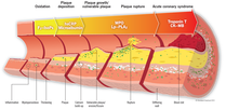
The purpose of this research is to identify a method of using computational simulations built from mathematical models as a tool in cardiovascular medicine. One particular example is the prevention and reduction of heart attacks and deaths due to vulnerable plaques caused by cardiovascular disease. To begin, an artery segment is idealized in order to create a generalized mathematical model. From this model, creation of a patient-specific model can begin by obtaining medical images. Given these images, imaging devices can generate stacks of cross sectional images in which the biological substances are represented in pixels as grey scale intensities at each slice. From identifying the areas of interest, extraneous features are removed from the computational model. With this, the segmented images can be used to automatically construct surface representations of certain geometric interest. Through a skeletalization process, a center line is identified along each branch; from here, arterial path lines are used to build a three-dimensional solid fluid computational model. This solid model contains regions that represent blood and the artery wall, and uses mesh resolution to adjust accurate boundary layers. Finally a solid NURBS mesh is constructed using isogeometric analysis (IGA) which in turns constitutes the fluid structure computational model for the system. This research could have a major breakthrough and turning point in the way clinical medicine is practiced, creating opportunities to better the lives of millions.
The purpose of this research is to identify a method of using computational simulations built from mathematical models as a tool in cardiovascular medicine. One particular example is the prevention and reduction of heart attacks and deaths due to vulnerable plaques caused by cardiovascular disease. To begin, an artery segment is idealized in order to create a generalized mathematical model. From this model, creation of a patient-specific model can begin by obtaining medical images. Given these images, imaging devices can generate stacks of cross sectional images in which the biological substances are represented in pixels as grey scale intensities at each slice. From identifying the areas of interest, extraneous features are removed from the computational model. With this, the segmented images can be used to automatically construct surface representations of certain geometric interest. Through a skeletalization process, a center line is identified along each branch; from here, arterial path lines are used to build a three-dimensional solid fluid computational model. This solid model contains regions that represent blood and the artery wall, and uses mesh resolution to adjust accurate boundary layers. Finally a solid NURBS mesh is constructed using isogeometric analysis (IGA) which in turns constitutes the fluid structure computational model for the system. This research could have a major breakthrough and turning point in the way clinical medicine is practiced, creating opportunities to better the lives of millions.
